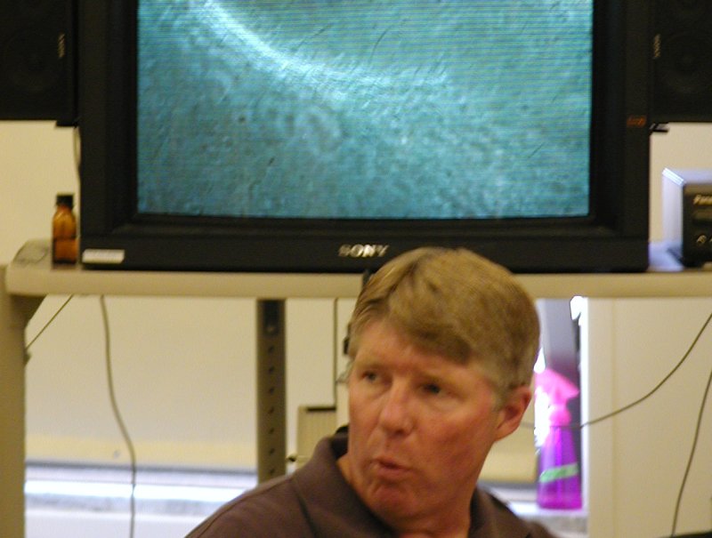
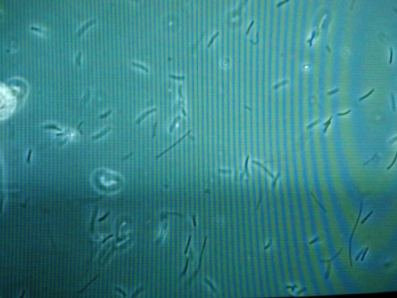
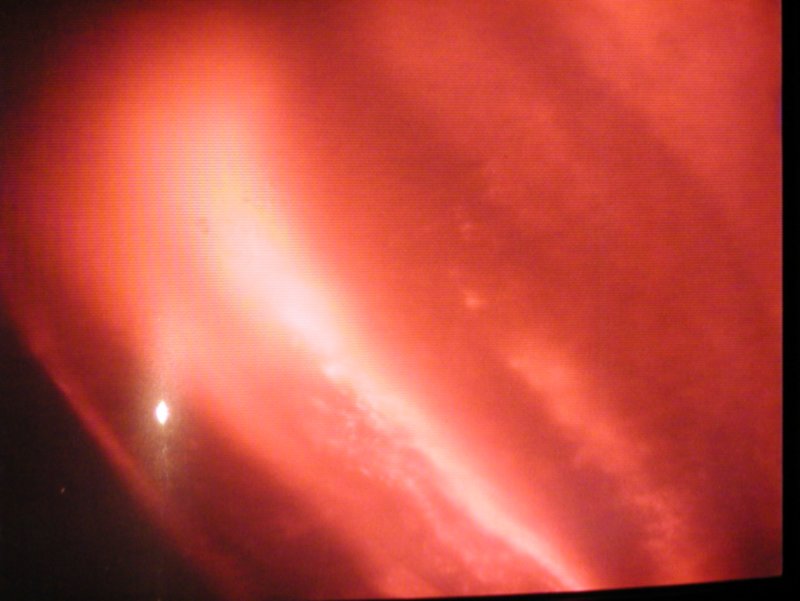
MICROBIOLOGY LABORATORY - 2 Back at Montana State University, Dr. Ward opened his teaching laboratory to us. He prepared the microbial mat samples we had collected for optical microscopy, and we saw what kinds of cells were in the mats. The colors on these images are not necessarily right, and the striping is from interference between the video
monitor and the digital camera and the overhead lights. |
Video microscopy lets the whole class see and discuss together! |
A pink tendril microbial goo from the very hot water (83°C) at Octopus Springs. The microbes here are Aquifex and Thermotoga which are the thin filaments all stuck together. Low magnification - the tendril is ~100 micrometers across, and the filaments are near a couple micrometers. |
|
Synechococcus cells and short filaments, from the green top of the Octopus Springs mat we collected. These filaments are about a micrometer across. |
Ultraviolet fluorescence of the bottom of the Octopus Springs mat we collected. Cyanobacteria fluoresce red and other types of microbes do not fluoresce. This view shows the complex structure of the microbial mat, with communities of funamentally with different organisms living interdependently in layers thinner than a millimeter. |