
MICROBIOLOGY LABORATORY - 1
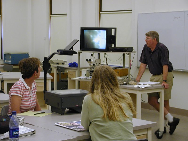
Back at Montana State University, Dr. Ward opened his teaching laboratory to us. He prepared the microbial mat samples we had collected for optical microscopy, and we saw what kinds of cells were in the mats.
|
|
|
MICROBIOLOGY LABORATORY - 1 Back at Montana State University, Dr. Ward opened his teaching laboratory to us. He prepared the microbial mat samples we had collected for optical microscopy, and we saw what kinds of cells were in the mats. |
 |
 |
|
Dr. Ward's teaching laboratory and microscope. The binocular scope has a video camera on the top (cables) that feed the TV above it on the cart. Joan and Lana watch raptly. |
Dr. Ward at the
microscope. Caption #1.
Caption #2.
|
|
 |
 |
|
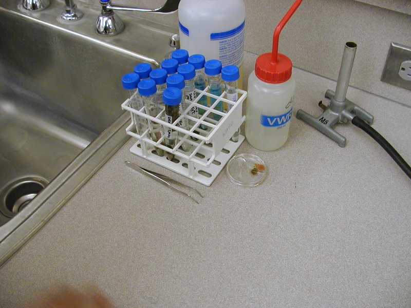
All of the biological and water samples we collected in Yellowstone Park. The blue tubes are sulfur-rich waters. The Petri dish contains the mat sample we collected at Octopus Springs. |
Dr. Ward teases a bit bacterial filament onto a glass slide for viewing in the microscope. The mat material goes in a shallow well on the slide, and is sealed in with a cover slip. This is the pink filament of Octopus Springs, from the hottest water there. |
|
|